Neuro Science
Neuroimaging is a technique for producing images of the human nervous system. It makes it possible to depict the anatomy or dynamic processes such as the flow of blood or cerebrospinal fluid.
Projects
- fMRI at 3T and 7T
- Diffusion-weighted MRI at 3T and 7T
- Magnetic resonance elastography
- Real-time-fMRI
- Human-machine-interactions in virtual realities
- Neuro- and biofeedback
- Equipment
- Cooperations
fMRI at 3T and 7T
Functional magnetic resonance imaging (fMRI) is a non-invasive imaging method that makes it possible to visualize the activity of nerve cells in the brain. It is widely used in neuroscientific research as well as in hospitals as a way of discovering more about the way the brain works. It uses what is known as the BOLD (blood oxygenation level-dependent) contrast method. Neurofeedback is a new area of application for functional MRI. This is a method with which the test subjects or patients receive information about their brain activity in real time, enabling them to focus on altering it.
Pharmaco-fMRI
Contact:
Publications:
Paper Gadolinium-enhanced magnetic resonance angiography in brain death
Paper Decreased effective connectivity in the visuomotor system after alcohol consumption
Paper Changes in gray matter volume after microsurgical lumbar discectomy
Paper Ethanol modulates the neurovascular coupling
fMRI for depth perception
Contact:
Publications:
This study examined the brain activity elicited by depth perception. As some studies had already identified that depth perception is lateralized to the right hemisphere, the sample size was increased to 26 test subjects in order to achieve a higher statistical significance. All subjects reported stable depth perception. In the random effects group analysis, the maximum activation where there was disparity in comparison with the condition without disparity was localized to a highly significant extent in the extrastriate cortex, presumably in V3A. The activation was greater in the right hemisphere. In the individual evaluations, however, a depth-related right hemisphere lateralization was only observed among 65% of the test subjects. The lateralization of depth by disparity may therefore be concealed in smaller groups.
Diffusion-weighted MRI at 3T and 7T
Contact: Dipl.-Ing. Ralf Lützkendorf
Basic Principles
DWI (Diffusion-Weighted Imaging) is used to map the motion of water molecules in the tissues. In strongly anisotropic tissues such as the white matter in the brain, nerve fiber orientations can be depicted through additional calculations such as DTI - Diffusion Tensor Imaging and HARDI - High Angular Diffusion Imaging. HARDI data are used to enable nerve fiber crossings and passes to be calculated, which, with models such as Fiber Orientation Distribution Function (fODF) or Track-Density Imaging (TDI) is able to solve such problems. What is known as fiber tracking is already established on 1.5 T magnetic resonance imaging (MRI) machines. By increasing the SNR at higher magnetic fields of 7 or more Tesla MRI in combination with stronger gradient fields of 70, 80 and mT/m2, in principle the spatial resolution and thus both the accuracy of the fiber orientations as well as the resolution of the functional measurements can be improved. Potentially, this raises expectations of new findings for functional and anatomically linked areas.
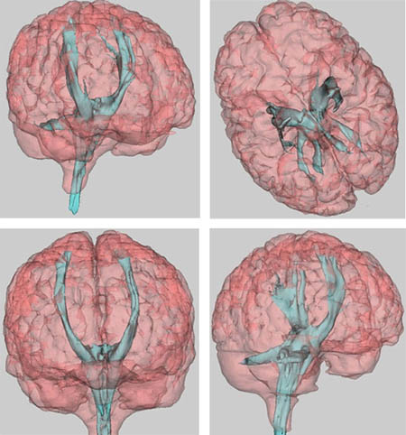
Volume rendering of the motor tract (light blue) IBMI 2006:
Track Density Imaging (TDI) of a sagittal layer calculated with MRTrix 3.0 IBMI 2015:
Fiber Orientation Distribution Function (fODF) calculated with MRTrix 0.2 (left: axial layer; right: coronal layer)
References (selected):
Lützkendorf, Ralf; Heidemann, Robin; Feiweier, Thorsten; Luchtmann, Michael; Baecke, Sebastian; Kaufmann, Jörn; Stadler, Jörg; Budinger, Eike; Bernarding, Johannes
Mapping fine-scale anatomy of gray matter, white matter, and trigeminal-root region applying spherical deconvolution to high-resolution 7-T diffusion MRI
In: Magnetic resonance materials in physics, biology and medicine - Heidelberg: Springer, vol. 31.2018, 6, pp. 701-713
Whole Paper Download:
Eichner, Cornelius; Setsompop, Kawin; Koopmans, Peter J.; Lützkendorf, Ralf; Norris, David G.; Turner, Robert; Wald, Lawrence L.; Heidemann, Robin
Slice accelerated diffusion-weighted imaging at ultra-high field strength
In: Magnetic resonance in medicine - New York, NY [u.a.]: Wiley-Liss, 1984, Bd. 71.2014, 4, S. 1518-1525
Eichner, Cornelius; Setsompop, Kawin; Koopmans, Peter J.; Anwander, Alfred; Lützkendorf, Ralf; Cauley, Steven; Bhat, Himanshu; Norris, David G.; Turner, Robert; Wald, Lawrence L.; Heidemann, Robin
Combining ZOOPPA and blipped CAIPIRINHA for highly accelerated diffusion weighted imaging at 7T and 3T
In: Discovery, innovation & application - advancing mr for improved health : ISMRM 21st Annual Meeting & Exhibition ; SMRT 22nd Annual Meeting Salt Lake City, Utah, USA 20-26 April 2013 ; programm, 2013, 2013, p. 0053
Lützkendorf, Ralf; Hertel, Frank; Heidemann, Robin; Thiel, Andreas; Luchtmann, Michael; Plaumann, Markus; Stadler, Jörg; Baecke, Sebastian; Bernarding, Johannes
Non-invasive high-resolution tracking of human neuronal pathways: diffusion tensor imaging at 7T with 1.2 mm isotropic voxel size
In: Medical imaging 2013: physics of medical imaging ; 11 - 14 February 2013, Lake Buena Vista, Florida, United States ; [part of SPIE medical engineering] / sponsored by SPIE. Robert M. Nishikawa ..., ed.: physics of medical imaging ; 11 - 14 February 2013, Lake Buena Vista, Florida, United States ; [part of SPIE medical engineering] - Bellingham, Wash.: SPIE, 2013; Nishikawa, Robert M., 2013, 866846, total of 7 p. - (Proceedings of SPIE; 8668) ; Kongress: Physics of medical imaging (Lake Buena Vista, Florida : 2013.02.11-14)
Publikationen:
E-Pub
Further scientific contributions of the research group:
R. M. Heidemann, A. Anwander, C. Eichner, R. Lützkendorf, T. Feiweier, T. R. Knösche, J. Bernarding, R. Turner: Isotropic sub-millimeter diffusion MRI in humans at 7T, 17th Annual Meeting of the Organization for Human Brain Mapping, Quebec City 2011, QC, Canada, 26 -30 May 2011.
R. Lützkendorf, R.M. Heidemann, A. Anwander, J. Stadler, T. Feiweier, O. Speck, J. Bernarding: In vivo DWI at 7T with a 70 mT/m gradient coil: 24- vs 32-channel head coil, 17th Annual Meeting of the Organization for Human Brain Mapping, Quebec City 2011, QC, Canada, 26 - 30 May 2011.
Hertel, Frank; Krefting, Dagmar; Lützkendorf, Ralf; Viezens, Fred; Thiel, Andreas; Peter, Kathrin; Bernarding, Johannes
Diffusion-Tensor-Imaging als Gridanwendung - Performanzsteigerung und standortunabhängiger Zugang zu leistungsfähigen Ressourcen
In: Informatik 2009. - Bonn : Ges. für Informatik, p. 116 - (GI-Edition) Kongress: Jahrestagung der Gesellschaft für Informatik e. V.; 39 (Luebeck) : 2009.09.28-10.02
Lützkendorf, Ralf; Bernarding, Johannes; Hertel, Frank; Viezens, Fred; Thiel, Andreas; Krefting, Dagmar
Enabling of Grid based diffusion tensor imaging using a workflow implementation of FSL
In: Healthgrid research, innovation and business case . - Amsterdam [u.a.] : IOS Press, ISBN 978-1-607-50027-8, pp. 72-81, 2009
Magnetic resonance elastography
Contact:
Magnetic resonance elastography (MRE) is an imaging technology for quantitatively mapping the characteristics of living tissue. This significantly differs from conventional methods such as fMRI, diffusion or proton density. MRE thereby fundamentally extends the diagnostic possibilities of magnetic resonance imaging (MRI).
Real-time fMRI
Contact: Dipl.-Ing. Sebastian Baecke
Publications and E-Pub
Projects
E-Pub
Hyperscanning
Real-time fMRI is based on the detection of brain activity using functional MRI. In this process, the aim is to analyze the functional data during an fMRI scan and to prepare the results for immediate use. Multivariate processes such as pattern-based classification processes increase the field of application of this method to include the analysis of complex brain patterns in real time.
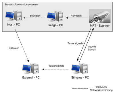
The illustration shows the technical structure of the implementation on the 3 and 7 Tesla MRI machines in the Faculty of Medicine at the University of Magdeburg.
Monitoring of brain activation in fMRI experiments
By using real-time fMRI it is possible to monitor the activation process even during an fMRI experiment and thus to detect sources of error that normally would only be noticed during post-experimental analysis.
Neurofeedback
Test subjects attempt to influence their brain activation based on feedback about this activation and thus achieve an impact on the behavioral mechanisms that triggered the state of activation.
Control of external systems
A computer is controlled solely using a test subject’s own brain activation. This allows activation patterns in imagined movements of limbs to be decoded and translated into control commands. As an example, this technology facilitates navigation in a virtual labyrinth.
Decision-making processes (Ultimatum Game)
Contact:
Publications:
Decision-making processes can be directly observed and evaluated using real-time MRI and real-time pattern analysis of brain activity. For more information read A new concept of a unified parameter management, experiment control, and data analysis in fMRI: Application to real-time fMRI at 3 T and 7 T and Predicting Decisions in Human Social Interactions Using Real-Time fMRI and Pattern Classification. Some members of the IBMI Neuro Science group participated in writing this paper.
BCI for navigation in virtual reality
Contact:
Publications:
--- under construction ---
BrainPong
Contact:
Publications:
--- under construction ---
Human-machine-interaction in virtual realities
Contact:
Publications:
E-Pub
Further scientific contributions of the research group:
S. Baecke: Ablauf, Datenaufkommen und Auswertung eines klassischen fMRT-Experiments, LABiMi/F Workshop Forschungsdatenmanagement in der medizinischen Bildverarbeitung, 29 February 2012, Magdeburg, Germany
S. Baecke: Implementierung eines Frameworks für das Hyperscanning, 42. Jahrestagung der Gesellschaft für Informatik, 16 - 21 September 2012, Braunschweig, Germany
S. Baecke: Echtzeit-fMRT und Neurofeedback, Symposium "Images and Networks of the Brain - New Methods and Perspectives, 24 - 25 September 2012, Hamburg, Germany
M. Lührs, C. Müller, J. Bernarding: A real-time fMRI communication interface for Turbo-BrainVoyager 3.0 using the TBV 3.0 Plugin Interface, 17th Annual Meeting of the Organization for Human Brain Mapping, 26 - 30 May 2011, Quebec City, QC, Canada
C. Hänel, C. Müller, J. Bernarding: Visualization and Modular Plug-In based Data- Analysis of Real-Time fMRI Data, 17th Annual Meeting of the Organization for Human Brain Mapping, 26 - 30 May 2011, Quebec City, QC, Canada
C. Müller, M. Lührs, S. Baecke, J. Bernarding: Stimulus application presenting virtual environments for real-time fMRI experiments, Joint annual meeting ESMRMB 2011, 6 - 8 October 2011, Leipzig, Germany
C. Müller, M. Lührs, A. Thoms, S. Baecke, J. Bernarding: A Qt-based virtual environment stimulus application for rfMRI using an improved TCP/IP framework, 17th Annual Meeting of the Organization for Human Brain Mapping, 26 - 30 May 2011,Quebec City, QC, Canada
A. Thoms, C. Müller, J. Bernarding: A brain-computer interface for navigation through a virtual environment in real-time fMRI studies, 17th Annual Meeting of the Organization for Human Brain Mapping, 26 - 30 May 2011, Quebec City, QC, Canada
C. Müller : A Human Brain Interface using a Virtual Environment System for real-time fMRI , German-Chinese Workshop on High-Field MRI, Hands-on Workshop at 7T, 26 - 27 April 2010, Magdeburg,Germany
C. Müller, M. Hollmann, N. Henniger, J . Bernarding: A virtual environment system for real-time fMRI (3T; 7T) using direct communication via TCP/IP, 16th Annual Meeting of the Organization for Human Brain Mapping, 6 - 10 June 2010, Barcelona, Spain
M. Hollmann, C. Müller, D. Adolf, J. Bernarding: Neural Correlates of social Decision-Making: Pattern-Classification of Bold-Responses during social, 16th Annual Meeting of the Organization for Human Brain Mapping, 6 - 10 June 2010, Barcelona, Spain
Brain-Computer interfaces
Contact: Dipl.-Ing. Sebastian Baecke
This project aims to design and implement a VR (Virtual Reality) stimulus environment for real-time fMRI studies. Virtual reality is a computer-simulated environment that conveys synthetic experiences to users. Thus, through this deeper visualization, everyday processes will be mapped more realistically and the subject's attention will be increased through more interaction with the paradigm.
The subject's brain activations within the motor cortex were measured and classified in an fMRI measurement. Based on these recorded brain activation patterns and an implemented decision algorithm, the subject was able to move an avatar through a virtual maze.
Use case: Realtime-fMRI Virtual Reality Experiment
Human-machine-emotion
Find out more about human-machine-interactions.
Virtual realities
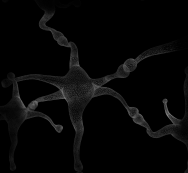
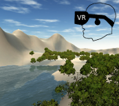
The VR group specializes in creating virtual realities (VR) on a wide variety of platforms and operating systems. The virtual environments are used in functional magnetic resonance imaging (fMRI), real-time fMRI (rtfMRI) or electroencephalography (EEG).
 |
 |
Contact: Dipl.-Ing. Ralf Lützkendorf
Completed projects:
Hardware and software:
Oculus Rift DK2
Oculus Rift Release
Microsoft Kinect 2
Unity3D
Unreal Engine
3D Studio Max
Blender
Poser 3D
Example:
BoldEffect_oculusRift_v01 (A walkthrough "Bold Effect" created with Unity3D for Oculus Rift DK2)
The BOLD effect (video):
Former team members:
B.Sc. Hannes Seibt
B.Sc. Julian Alpers
Marina Knaus
Alexander Roewer
Gerd Schmidt
Svenja Völler
Adaptive brain-computer interface in stress paradigms
Publications:
Automatic Adaptation of Stimulus to Stress Level (Emoadapt)
In the EMOADAPT project, the environment or task of the test subject is adapted to their mood or stress level. To carry out the test, a program was developed with a dexterity task that the test subject could continuously complete during fMRI measurement. (Blocky).
The level of difficulty of the dexterity test can be continuously adjusted in the process, while at the same time the stress intensity of the test subject is almost continuously determined virtually from the constantly updated BOLD signal images from the fMRI measurements (BCI, brain computer interface).
For the practical implementation of this idea, the question arises as to how much a difference between the target and actual test subject stress should increase or decrease the difficulty of the dexterity task. This question can be answered using classic control theory methods.
Control loop for adapting the difficulty of the dexterity task

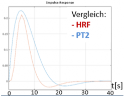
A step change in the task difficulty leads to a stepped response in the resulting brain functions. However, for the measured BOLD signal, there is a distortion and delay that is usually described by convolution with the hemodynamic response function (HRF). This is modeled as a PT2 element in the context of the control system description. A comparison of the HRF convolution kernel and the PT2 convolution kernel is illustrated below:
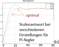
From the model, the parameters can then be calculated that lead to the level of difficulty of the dexterity task being corrected in such a way that there is neither an overmodulation with many subsequent overshoots nor an unnecessarily slow convergence on the optimum value.
Multivariate real-time fMRI decoding
Contact: Dirk Schomburg
Publications:
D. Schomburg 2018, Ansätze für die funktionelle Magnetresonanztomographie-Dekodierung in Echtzeit mit multivariaten Methoden: Master thesis
D. Schomburg, M . Plaumann, J. Bernarding 2020, Approaches for real-time fMRI decoding using multivariate methods; GMDS2020
Multivariate approaches to real-time fMRI decoding can improve the accuracy of the decoding of brain functions from the current BOLD signal volume images. As an introduction to the subject, it is first necessary to understand the concept of fMRI and real-time fMRI decoding. These are briefly explained in the following paragraphs.
Introduction
Functional magnetic resonance imaging (fMRI) makes it possible to detect different areas of the brain that are more strongly activated by stimuli or mental tasks. fMRI works on the basis of the different magnetic characteristics of blood before and after oxygen consumption resulting from neuron activity. The measured signal is termed the blood oxygen level-dependent (BOLD) signal.
With real-time fMRI decoding, conclusions can be drawn about the current brain function from the knowledge of the conditionally anticipated more strongly activated brain areas and the spatially-resolved measured BOLD signal. An fMRI pre-measurement is generally used as a training phase for the parameters used to adjust the prediction functions for fMRI decoding. A simple possibility for this is the use of masks of significant voxels from the fMRI pre-measurement. If the average value of the BOLD signal in the masks in the current volume image is calculated, this results in a rough decoding of the current brain function.
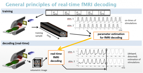
Decoding should be improved with multivariate methods so that decoding is possible even with weak neuronal activities. Some of the challenges in this respect are as follows:
- Due to the large number of voxels (>150,000) and the small sample size (100-500) it is not possible to obtain a predictor using the unmodified classic regression technique.
- Modern regularized regression methods use an inverse model whose random errors cannot be interpreted. Apart from this, the necessary determination of the hyper parameters leads to a strong increase in the training computation effort.
- In addition, the measured values are often overlain by a random trend for which linear modeling is not adequate. Conventional trend adjustment methods use the entire measurement time series; this is only possible here in the training phase, while real-time decoding requires trend adjustment that only requires the measured values acquired up to the time of measurement.
Methods
In simulation calculations, the computing time and prediction risk for various scenarios were calculated and compared for each of the ad-hoc method, a stabilized classic method, a regularized modern method and for principle component regression.
To take trends into account, the model was supplemented with a Gaussian random walk. Restricted maximum likelihood estimators yield the variance components. Thus, pre-whitening the data leads to data with homoscedastic errors for which the regression methods are applicable. The predictors with the model parameters obtained in this way produce trended time series, whose trend adjustment became possible with additional prior knowledge of the stimulation time series.
Results
The principle component regression method was shown here to be the best compromise between training computing time and prediction risk. It was possible to find trend consideration methods for training and decoding. Simulated and measured data showed good results for the approach that was developed for fMRI decoding.
Decoded time series with the simple average method

Decoded time series from the same data but with the multivariate method, which is based on principle component regression

Extension
By improving the algorithms, it was possible to ensure that the method described above could be applied not only to classification tasks bus also to block paradigms with different intensities in the blocks.
Decoded time series with the simple average method for different intensities in the blocks:

Decoded time series from the same data, but with the multivariate method, which is based on principle component regression and was adjusted for different intensities in the blocks.

Realt-time pipeline between BCI and VR or feedback systems
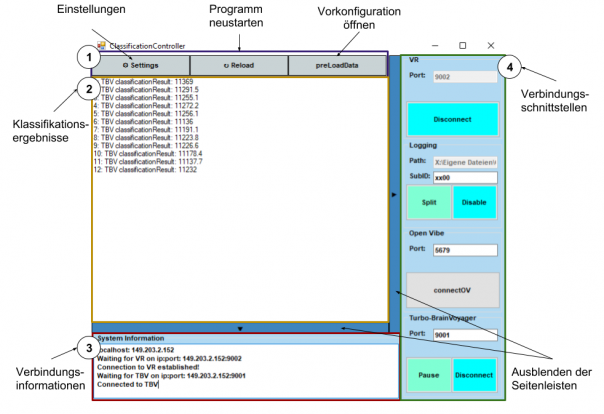
Current individual measured values or measured value vectors are output at short intervals from the brain computer interface (BCI). These are then to be used to control virtual reality (VR), other feedback representations or other systems. A TCP/IP interface has been developed at the Institute for the transmission of data between the components, which also enables the transmission of data to remote computers.
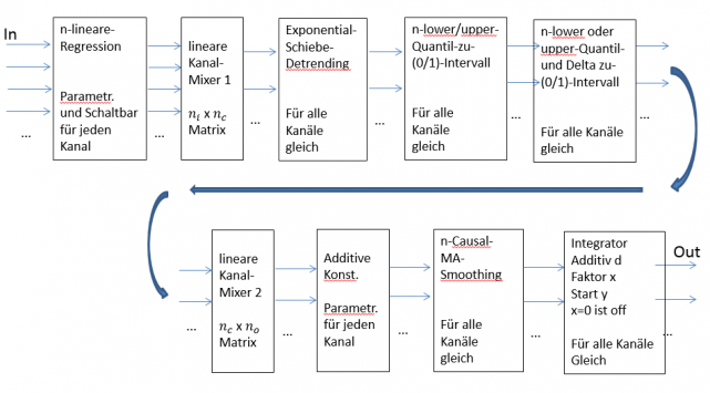
Often, this signal transmission requires additional adjustments and calculations. In the simplest case, this would be a level adjustment and scaling to convert values between 1200 and 1300 to a signal between 0 and 1. Complicated tasks include signal smoothing functions, mixing of several channels or elements from control technology (PID controllers).
A log function is required for troubleshooting and parameter adjustment and status displays and control functions should be easily accessible for the TCP/IP interfaces. To be able to quickly use these during real-time fMRI experiments, these functions were implemented in a software package with a graphical user interface.
Configurable signal routing function blocks:
Graphical user interface
Neuro- and biofeedback
This work implemented a brain-computer interface to improve human-machine interaction. The aim was to enable the adaptation of a virtual environment in the form of a game to the state of an operator, which was detectable by means of EEG and biophysical data, and to adaptively regulate any difficulty. To realize this task, a preliminary study was evaluated and based on this a study was planned and conducted that measured the seventeen test subjects multiple times. The study involved plotting both a psychological standard paradigm and a VR game scenario. This study took place in a real, unshielded working environment in order to cover the real needs of use cases. The data were evaluated and transformed into a suitable feature vector. Based on this vector, two artificial neural networks were created, their structures were compared, and different parameters were evaluated in an offline analysis. In the process it was demonstrated that the developed feature vector could be used for the classification of both standard paradigms and also the different difficulty levels of the game used. The trained neural networks could then be loaded into a network-based data processing pipeline created for this project, so that it could classify EEG and biophysical data in real time. Within this pipeline, various important modules for real time data capture and processing as well as communication via network were designed and implemented.
Equipment
Laboratory for the Development of Real-Time Virtual Reality and Pattern Recognition (EEG, bio-psychological parameters)
Facilities include 2 portable EEG machines for use in bio- and neurofeedback (4 channels), 2 MRI-compatible EEG machines (64 channels), one transcranial stimulation device (TCD), 2 VR headsets (Oculus Rift), one AR headset (HoloLens 2) and the corresponding software (commercial and proprietary).
The institute can measure on the following whole-body MRI systems:
- Unity3D
- Unreal Engine
- Oculus Rift DK2
- Kinect Microsoft
- 3-Tesla-Magnetresonanz-Tomograph (Siemens MAGNETOM Prisma (2015)) (Betreiber: Universiätsklinik für Neurologie)
- 7-Tesla-Magnetresonanz-Tomograph (Siemens MAGNETOM) (Betreiber: LIN)
Cooperations
Cooperation partners
- Prof. Dr. Ingolf Sack, PD Dr. Jürgen Braun (AG Elastographie, Charité Berlin)
- Prof. Dr. Felix Blankenburg (FB Psychologie, FU Berlin)
- Leibniz-Institut für Neurobiologie (LIN) Magdeburg
- Prof. Dr. Oliver Speck (Fakultät für Physik, Lehrstuhl Biomedizinische Magnetresonanz, OvGU)


